
产品名称:Anti-TNF Antibody, Rabbit Polyclonal
经验证的应用:WB/IHC
交叉反应:Predict reacts with: Human/Mouse/Rat
特异性:mouse TNF
免疫原:Recombinant mouse TNF protein, fragment Leu80~Leu235; UniprotKB: P06804
制备方法:Produced in rabbits immunized with mouse TNF, and purified by antigen affinity chromatography.
来源:Polyclonal Rabbit IgG
纯化:Immunogen affinity purified
缓冲液:Supplied in PBS, 50% glycerol and less than 0.02% sodium azide, PH7.4
偶联物:Unconjugated
状态:Liquid
运输方式:This antibody is shipped as liquid solution at ambient temperature. Upon receipt, store it immediately at the temperature recommended.
储存条件:This antibody can be stored at 2℃-8℃ for one month without detectable loss of activity. Antibody products are stable for twelve months from date of receipt when stored at -20℃ to -80℃. Preservative-Free. Avoid repeated freeze-thaw cycles.
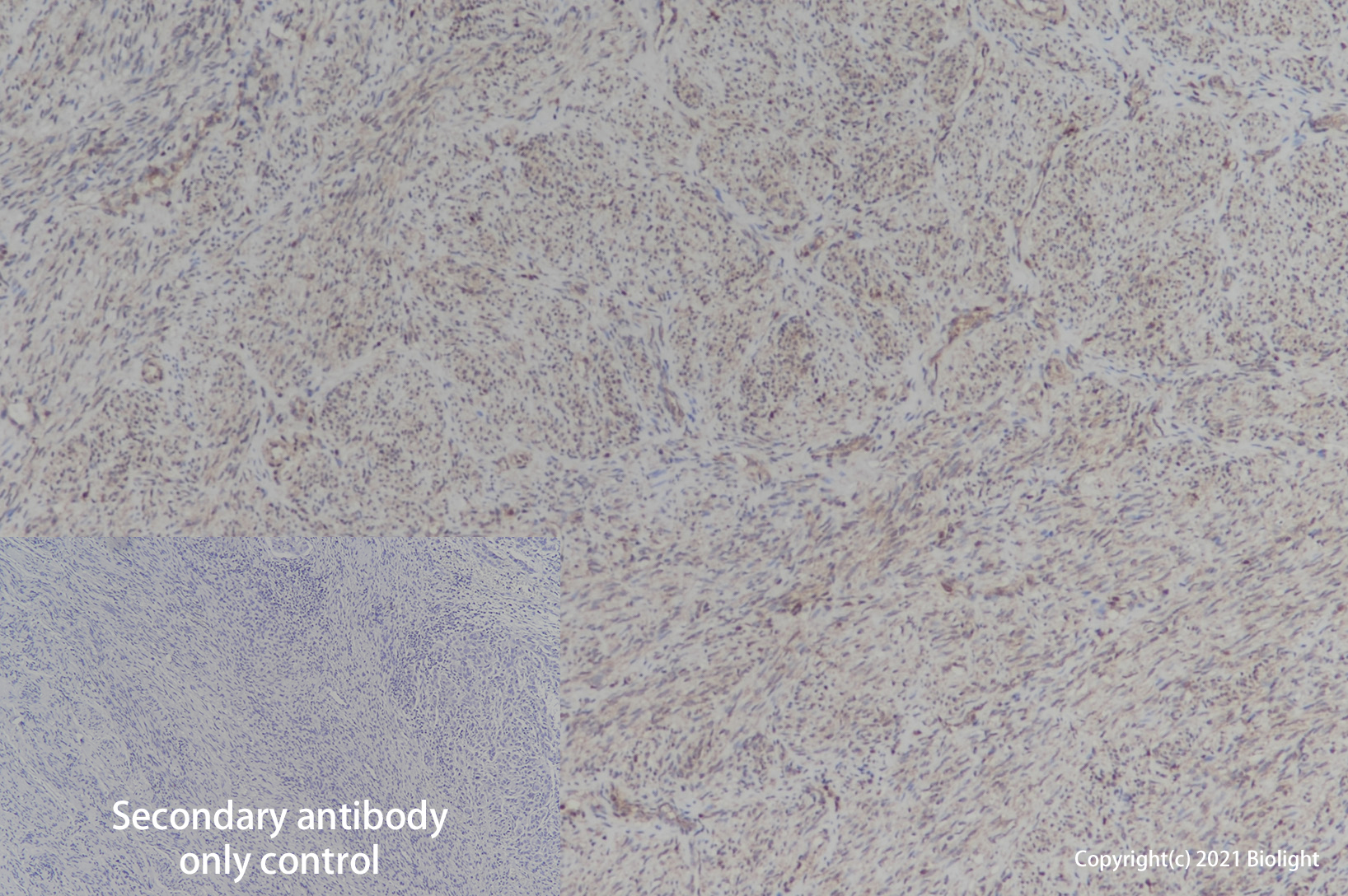
图片:
Figure1.Application in WB
Figure2.Application in IHC
别称:DIF, TNF-A, TNFSF2, Cachectin, Tumor Necrosis Factor Ligand Superfamily Member 2
背景信息:TNF-alpha. Tumor necrosis factor alpha (TNF-alpha ), also known as cachectin and TNFSF2, is the prototypic ligand of the TNF superfamily. It is a pleiotropic molecule that plays a central role in inflammation, immune system development, apoptosis, and lipid metabolism (1, 2). Bovine TNF-alpha consisits of a 35 amino acid (aa) cytoplasmic domain, a 21 aa transmembrane segment, and a 178 aa extracellular domain (ECD) (3). Within the ECD, bovine TNF-alpha shares 64%-83% sequence identity with canine, cotton rat, equine, feline, human, mouse, porcine, rat, and rhesus TNF-alpha. TNF-alpha is produced by a wide variety of immune, epithelial, endothelial, and tumor cells (1, 2). TNF-alpha is assembled intracellularly to form a noncovalently linked homotrimer which is expressed on the cell surface (4). Cell surface TNF-alpha can induce the lysis of neighboring tumor cells and virus infected cells, and it can generate its own downstream cell signaling following ligation by soluble TNFR I (2, 5). Shedding of membrane bound TNF-alpha by TACE/ADAM17 releases the bioactive cytokine, a 55 kDa soluble trimer of the TNF-alpha extracellular domain (6-8). TNF-alpha binds the ubiquitous 55-60 kDa TNF RI (9, 10) and the hematopoietic cell-restricted 80 kDa TNF RII (11, 12), both of which are also expressed as homotrimers (1, 2, 13). Both type I and type II receptors bind TNF-alpha with comparable affinity (14), although only TNF RI contains a cytoplasmic death domain which triggers the activation of apoptosis. Soluble forms of both types of receptors are released and can neutralize the biological activity of TNF-alpha (15).
全称:Tumor necrosis factor (TNF)